
Recent research published in journals Frontiers of nutrition evaluated the effects of dietary vitamin A on the gut microbiota and gut transcriptome.
Alzheimer’s disease (AD) is an age-related neurodegenerative disease. A 2021 report reveals that more than 55 million people worldwide are living with dementia, with low- and middle-income countries accounting for an estimated 60% of the total. Given the aging of the population, the prevalence of Alzheimer’s disease and its associated economic burden are expected to increase. Currently, no treatments exist that can cure Alzheimer’s disease or modify its pathological progression.
Therefore, effective management and regulation of related factors has become of paramount importance. Diet has been implicated in the prevention and progression of Alzheimer’s disease, and vitamin A is a potential prevention and treatment strategy. Non-genomic effects of vitamin A in the brain have important roles, and modulating them may improve brain function and provide therapeutic avenues.
 Study: Dietary vitamin A alters the gut microbiota and intestinal tissue transcriptome, affecting intestinal permeability and release of inflammatory factors, thereby influencing Aβ pathology. Image credit: Nefedova Tanya / Shutterstock
About research
In this study, researchers investigated the effects of dietary vitamin A on the intestinal transcriptome, inflammation, gut microbiota, and amyloid-β (Aβ) pathology. He randomly assigned 30 APP/PS1 mice (AD mouse model) into three groups based on body weight. Mice were fed diets with vitamin A-deficient (VAD), normal (VAN), or enriched (VAS) levels. Body weight and food intake were recorded weekly.
Animals were euthanized after 12 weeks. Serum and fecal samples were collected. Tissue was taken from the brain and intestines. Using enzymes to measure serum levels of intestinal permeability markers such as D-lactate and diamine oxidase (DAO), and cytokines such as interleukin (IL)-1β, IL-6, and tumor necrosis factor (TNF)-α. I measured it. Linked immunosorbent assay (ELISA). Neurobehavioral function was assessed with a 6-day Morris water maze test.
Therefore, mice were trained for 4 days and then explored the hidden platform for 60 seconds. After 5 days of training, a detection test was performed to determine the time to find the platform. On the last day he had two exams conducted. One required locating the platform within 60 seconds if it was not present, and the other measured the time it took to locate the platform.
Additionally, morphometric and immunohistochemical analyzes were performed to assess Aβ deposition in the brain. To analyze the gut microbiota, 16S rRNA sequencing was performed. Transcriptome analysis of intestinal tissue was also performed. Serum vitamin A levels were quantified by liquid chromatography-mass spectrometry.
Investigation result
There were no significant changes in body weight and food intake between groups throughout the intervention. However, at different time points, the VAS and VAD groups had lower body weight and food intake than his VAN group. The mean vitamin A level was 382.61 ng/ml in the VAD group, 548.32 ng/ml in the VAN group, and 640.85 ng/ml in the VAS group. Analysis of variance showed significant differences between groups.
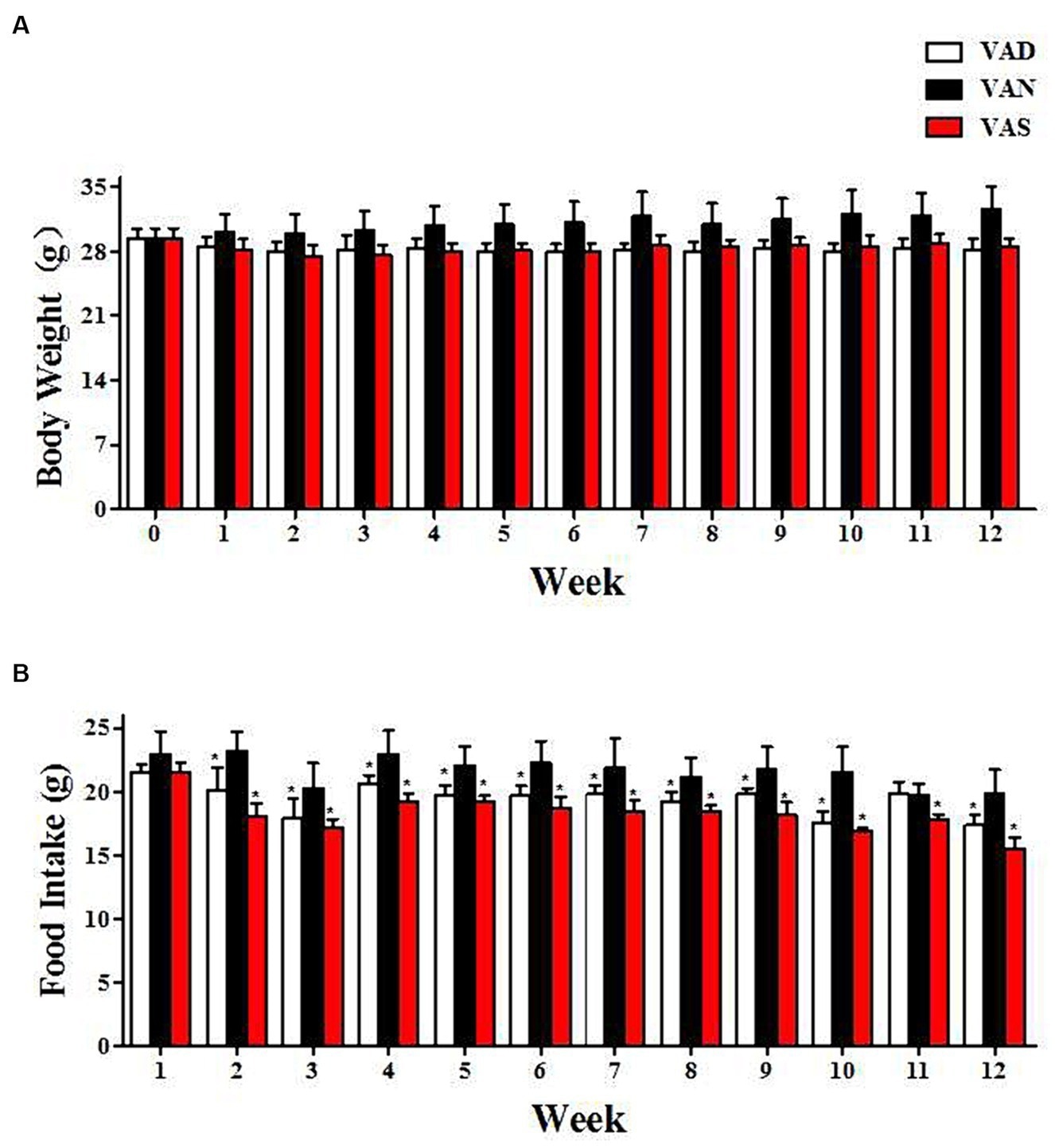
Effect of vitamin A on dietary intake and body weight changes in APP/PS1 mice. (A) Changes in body weight of APP/PS1 mice between different groups. (B) Changes in dietary vitamin A intake between different groups of APP/PS1 mice. *Indicates comparison with VAN group, p < 0.05 indicates statistical significance
Mice in the VAN group were able to directly locate the platform and move to its position even if removed. These mice took less time to find the platform than the other groups. Furthermore, the VAS group took less time than the VAD group. There was no significant difference in the time spent finding the platform between the VAS and VAN groups. After the platform was removed, the VAN group crossed the area many times.
Mice in the VAD group showed significantly more Aβ deposits in the brain than other groups. The VAS group also had more she Aβ deposits than his VAN group, although it was not statistically significant. Shannon index was higher in the VAS and VAN groups than in his VAD group. however, it was similar between the VAS and VAN groups. The VAN and VAS groups had better microbial diversity than the VAD group.
Overall, 571 differentially expressed genes were identified between the VAS and VAD groups. 313 differentially expressed genes were identified between the VAN and VAD groups. Furthermore, there were 243 differentially expressed genes between the VAN and VAS groups. DAO and D-lactate levels were significantly elevated in the VAD group compared to the VAS and VAN groups. Mice in the VAD group showed significantly higher levels of inflammatory cytokines than other groups.
conclusion
In summary, this study showed that a 12-week VAD diet reduced serum retinol levels, impaired cognitive function, and increased Aβ pathology in mice. Conversely, a diet rich in vitamin A increased retinol, decreased Aβ, and preserved cognition. Overall, this finding highlights the importance of vitamin A in Alzheimer’s disease pathology and behavior.
Reference magazines:
- Wang ZL, Pang SJ, Zhang KW, Li PY, Li PG, Yang C. Dietary vitamin A alters the gut microbiota and intestinal tissue transcriptome, leading to intestinal permeability and release of inflammatory factors. and thereby affect Aβ pathology. front nut2024, DOI: 10.3389/fnut.2024.1367086, https://www.frontiersin.org/articles/10.3389/fnut.2024.1367086/full